Debris, Particles and Fibres in Food, Drink and Pharmaceuticals
 LPD Lab Services Limited have two decades of experience in isolating and analysing foreign particles in foods, beverages, pharmaceutical products, clinical products, medical devices and formulations.
Often visible particles or fibres in solution are identified by the customer by eye or using a reliable liquid inspection system such as the LV28 This employs a cross-polarisation technique that may easily illuminate particles within solutions, where the particles scatter light and, therefore, appear to glow brightly against a dark background. The lab cross-polariser system allows staff to identify and isolate of particles from clear liquids for further in-depth analysis using techniques involving optical microscopy, scanning electron microscopy SEM / EDX, and either macroscopic Fourier transform infra-red FTIR spectroscopy or a state-of-the-art microATR FTIR microscope depending on whether contaminants are inorganic or organic in nature.
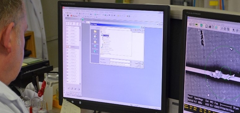
 Particles are usually isolated and cleaned as required, then transferred to a suitable substrate, like a glass slide or sticky SEM stub, and then overviewed in colour images under the optical microscope before transferring to the SEM / EDX for analysis.
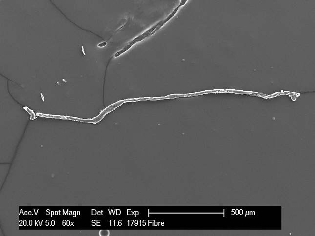
For more detailed analysis of the particles in the SEM using both secondary electron SE and backscatter electron BSE imaging modes.
- The SE greyscale images provide topographic information and are used to look at the size and morphology of the particle or fibres found to help indicate the origin of the particles like wear particles, natural or synthetic fibres.
- The BSE greyscale images provide information about the chemical uniformity of the particles surface with a uniform grey shade indicating a uniform elemental composition. Whiter areas in a BSE image indicate the presence of higher atomic number elements, such as sub-micron metallic particles on an organic fibre. Darker areas may be indicative lower atomic number elements typical of organics like natural organic materials or synthetic polymers or rubbers. Areas showing different shades of grey may be analysed for their elemental signature by energy dispersive X-ray EDX analysis, particularly for inorganic or metallic contaminants. Areas high in carbon and oxygen are indicative of organic materials which may be further explored using either macroscopic Fourier transform infra-red FTIR spectroscopy or a state-of-the-art FTIR microscope.
Larger organic particles (>1 mm across) may be explored using macroscopic FTIR spectroscopy using either a diamond or germanium ATR, while smaller organic particles and fibres (<1 mm across) may be explored using a state-of-the-art FTIR microscope fitted with a germanium micro-ATR prism down to 20micrometres.