
SEM Principle and Instruments
 Principle of SEM / EDX
An SEM is essentially a high magnification microscope, which uses a focussed scanned electron beam to produce images of the sample, both top-down and, with the necessary sample preparation, cross-sections. The primary electron beam interacts with the sample in a number of key ways:-
- Primary electrons generate low energy secondary electrons, which tend to emphasise the topographic nature of the specimen.
- Primary electrons can be backscattered which produces images with a high degree of atomic number (Z) contrast.
- Ionized atoms can relax by electron shell-to-shell transitions, which lead to either X-ray emission or Auger electron ejection. The X-rays emitted are characteristic of the elements in the top few μm of the sample and are measured by the EDX detector.
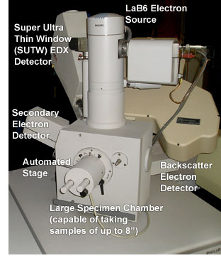
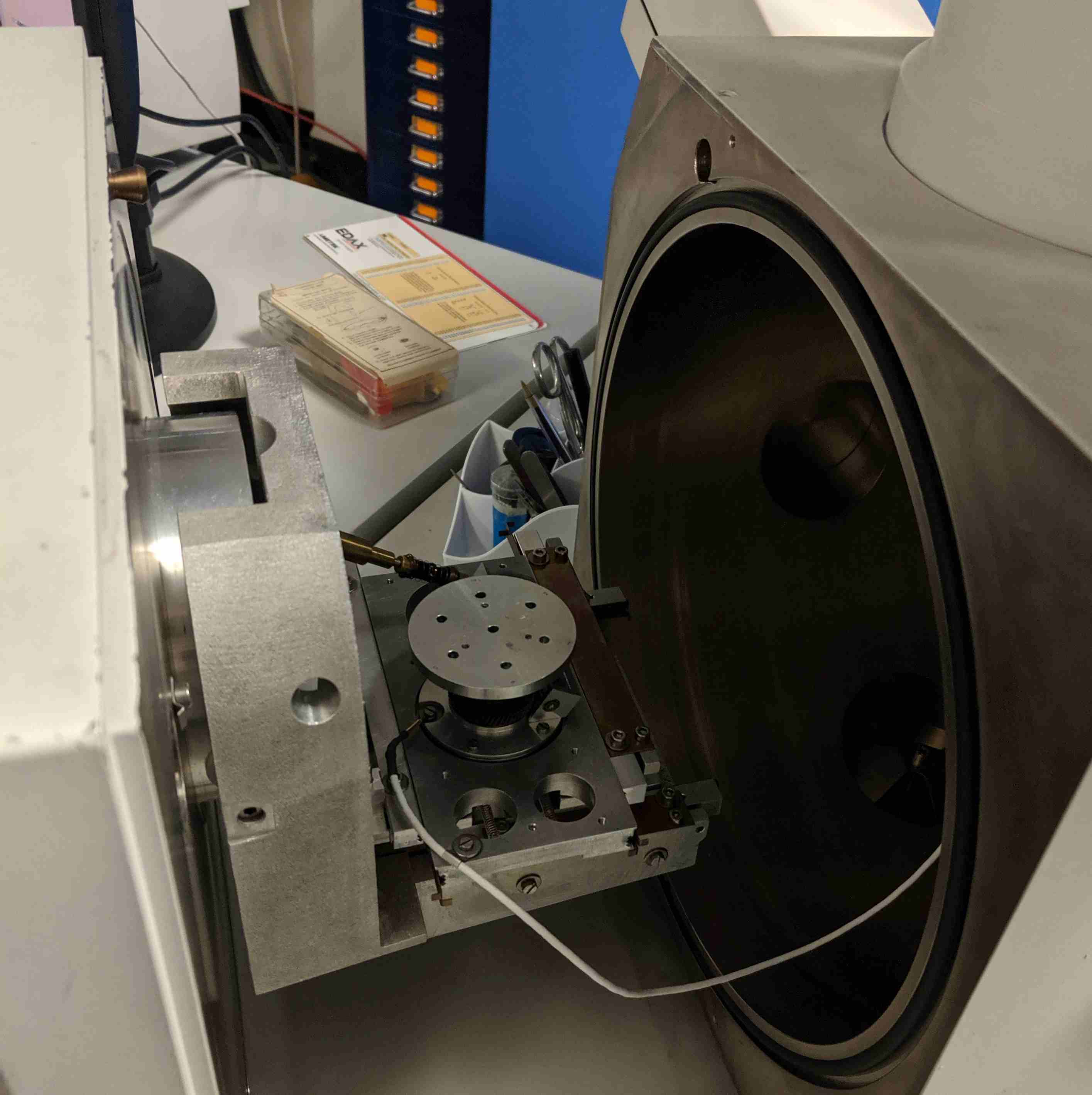
Our SEM / EDX Instruments
The scanning electron microscope is an instrument used for the imaging and analysis of a wide range of materials in a wide range of applications. The laboratory has 2 such instruments in house, one with a LaB6 tip and one with a tungsten filament, and 5 very experienced SEM analytical scientists. Additionally the company has access to higher resolution FEG (Field Emission Gun) instruments and environmental SEMs at trusted partner laboratories when required.
SEM Instruments - Philips XL30 Scanning Electron Microscopes and EDAX
 The main features and benefits of the SEM are:-
- Image magnification and resolution
- Magnification range X 15 - X 200,000
- Resolution 2 nm
- Accelerating voltage 1 - 30 keV
- Secondary and backscatter electron imaging
- Stereo imaging and stereo height measurement
- EDX analysis of known or unknown materials (www.edax.com)
- Qualitative and quantitative analysis for all elements from carbon upwards
- Quantitative analysis of bulk materials and features ≥ 2 μm
- Qualitative analysis of features ≥ 0.2 μm
- Detection limits typically 0.1 - 100 Wt% for most elements
- Multi-element X-ray mapping and line scans
- Multi-layer, multi-element thin film analysis - Thickness and composition
- Particle / Phase analysis - Detection, analysis, morphology and size
- Image Analysis
- Automatic particle counting and characterisation
- Large samples can be analysed up to half a house brick size.
Links
UK Institute of Physics Electron Microscopy & Analysis Group (MAG): http://groups.iop.org/EM/
Museum of Science- How an SEM Works www.mos.org/sln/sem/intro.html